
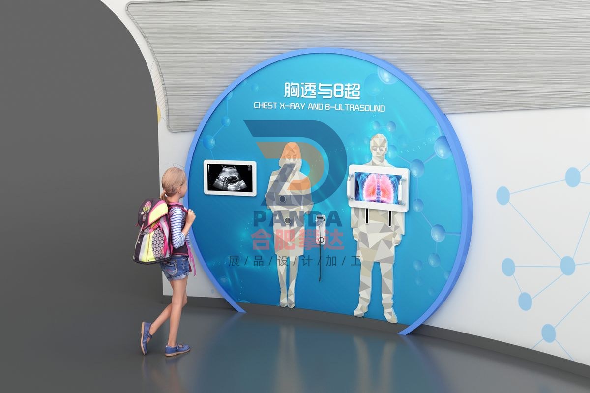
B-ultrasound and chest X-ray
Health & Wellness · Interactive
- DimensionsSubject to on-site plan
- PresentationInteractive
- PricingQuoted by manufacture spec
B-ultrasound and chest X-ray is a hands-on, mechanical & electromechanical interactive exhibit manufactured by Hefei Panda for science centers, children’s museums, discovery centers, STEM learning centers and family entertainment centers. Engineered for durable, robust, low-maintenance, high-traffic gallery installation, it turns abstract scientific concepts into direct, interactive visitor exploration. Hefei Panda is a global exhibit manufacturer exporting hands-on science displays to 50+ countries, with in-house engineering, fabrication, installation and commissioning.
Exhibit Summary
B-ultrasound and chest X-ray is a Interactive interactive exhibit in the Health & Wellness category, designed for science centers, children’s museums, discovery centers, STEM learning centers and family entertainment centers. Built as a hands-on, mechanical & electromechanical display, it turns an abstract scientific concept into direct, interactive visitor exploration, and is engineered for durable, low-maintenance, high-traffic gallery use.
Scientific Principle
Through interaction, let participants grasp the methods and indications of chest X-ray and B-ultrasound examination, and also teach participants how to watch.
Operation
This exhibition includes two sets of human body models for electromechanical interaction, which are used to show B-ultrasound examination and chest X-ray examination respectively. B-ultrasound examination: It includes a human silhouette board, a display screen and a detector head, etc. Guests can watch the B-ultrasound images via interaction. The function of playing B-ultrasound during standby and the impact of radiation. Chest X-ray examination: It includes a human silhouette board and a sliding screen. Guests can watch the real-time picture of the chest X-ray examination via interaction. Chest X-ray, called fluoroscopy, is a commonly used X-ray examination method. It makes use of the penetrability, fluorescence and photographic effects of X-rays to form an image of the human body on the screen. Since human tissues have differences in density and thickness, when X-rays penetrate different tissues of the human body, the X-rays are absorbed to different degrees, so the amount of X-rays reaching the screen is different, forming images with different black and white contrasts, providing a basis for doctors' diagnosis. B-mode ultrasound is the main ultrasound examination method, and its scope is very wide. Different examination parts require different preparations before examination.
Export-Ready Features
- Durable, robust construction for high-traffic gallery environments
- Low-maintenance electromechanical design with reliable, safe components
- Custom sizing, theming & multilingual labeling available
- OEM & turnkey exhibit solutions with full project management
- Overseas shipping, export documentation & on-site installation supported
- Shipped to science centers and museums in 50+ countries