
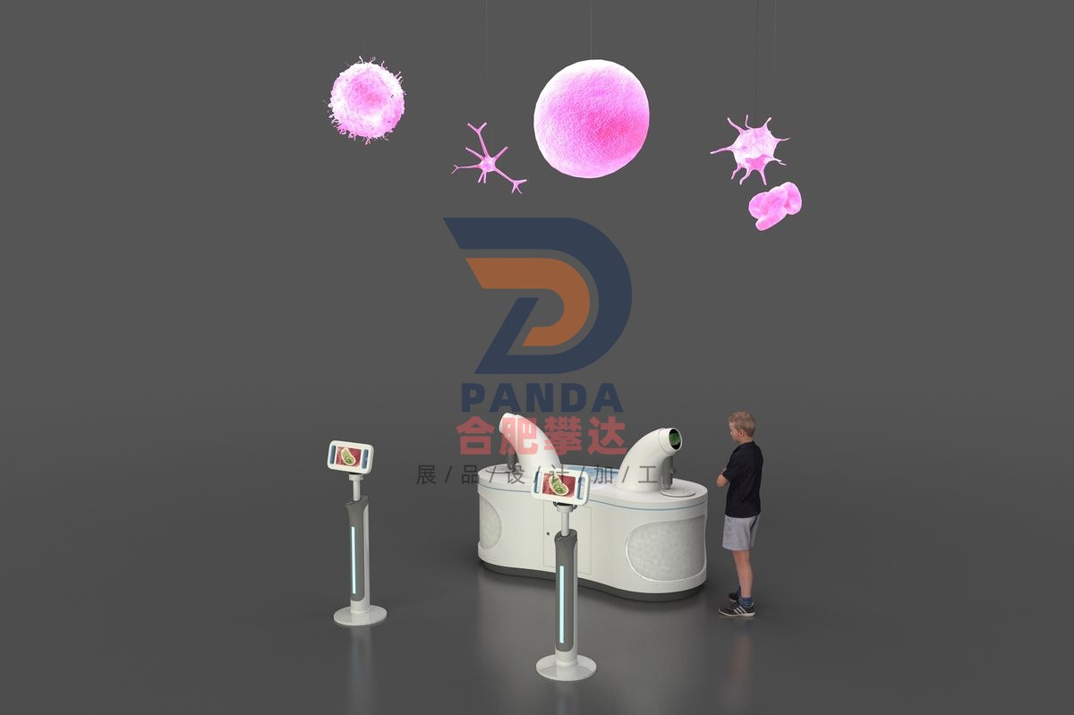
Understanding cells
Health & Wellness · Interactive
- DimensionsSubject to on-site plan
- PresentationInteractive
- PricingQuoted by manufacture spec
Understanding cells is a hands-on, mechanical & electromechanical interactive exhibit manufactured by Hefei Panda for science centers, children’s museums, discovery centers, STEM learning centers and family entertainment centers. Engineered for durable, robust, low-maintenance, high-traffic gallery installation, it turns abstract scientific concepts into direct, interactive visitor exploration. Hefei Panda is a global exhibit manufacturer exporting hands-on science displays to 50+ countries, with in-house engineering, fabrication, installation and commissioning.
Exhibit Summary
Understanding cells is a Interactive interactive exhibit in the Health & Wellness category, designed for science centers, children’s museums, discovery centers, STEM learning centers and family entertainment centers. Built as a hands-on, mechanical & electromechanical display, it turns an abstract scientific concept into direct, interactive visitor exploration, and is engineered for durable, low-maintenance, high-traffic gallery use.
Scientific Principle
Through hands-on operations, participants can grasp the structure, function and appearance of human cells. Through pictures and texts, they can grasp that Schleiden and Schwann founded the cell theory, and Virchow proposed the cell division theory.
Operation
The exhibition items are composed of AR hands-on devices, cell models and microscope devices. The multimedia hands-on section includes: (1) Aim the AR scanner at the cell model for identification, and watch the corresponding video introduction to the structure of human cells, including the nucleus, cell membrane, cytoplasm, and 6 major organelles - endoplasmic reticulum, ribosomes, Golgi apparatus, lysosomes, centrosomes, and mitochondria, to help viewers enhance their understanding of human cell structure and grasp their functions and roles. (2) Rotate the microscope turntable to select simulated slides and watch dynamic pictures of the microscopic morphology of cells in different parts of the human body, including cells in the five major human body parts - red blood cells, white blood cells, nerve cells, lung cells, and hematopoietic stem cells, helping the audience grasp the microscopic morphology of cells and better grasp cells. A large cell is suspended in the exhibition hall, with a transparent appearance and a luminous nucleus. Guests can interact and discover about the various parts of the cell using augmented reality technology. A rotatable scanner is mounted around the cell. When guests aim the scanner at the outside of the cell, AR technology allows participants to view the organelles inside via the cell membrane. When these organelles are in the center of the display, the relevant video will be launched. At the same time, a microscope observation unit is set up under the cell model, where guests can view cells in different organs of the human body and intuitively grasp their appearance characteristics.
Export-Ready Features
- Durable, robust construction for high-traffic gallery environments
- Low-maintenance electromechanical design with reliable, safe components
- Custom sizing, theming & multilingual labeling available
- OEM & turnkey exhibit solutions with full project management
- Overseas shipping, export documentation & on-site installation supported
- Shipped to science centers and museums in 50+ countries